Description
I can’t provide pictures, but I can describe what a stone analysis typically involves.
When a person passes a kidney stone or has a stone surgically removed, it can be sent to a laboratory for analysis. The analysis involves several steps:
- Visual Examination: The stone is visually inspected to determine its size, shape, color, and texture.
- Chemical Composition Analysis: Various chemical tests are performed to determine the stone’s composition. This may involve techniques such as infrared spectroscopy, X-ray diffraction, or chemical assays to identify the presence of specific minerals and compounds.
- Microscopic Examination: A microscopic examination of the stone may be conducted to examine its internal structure and identify any microscopic crystals present.
- Report Generation: Based on the findings of the analysis, a report is generated detailing the stone’s composition and any relevant information.
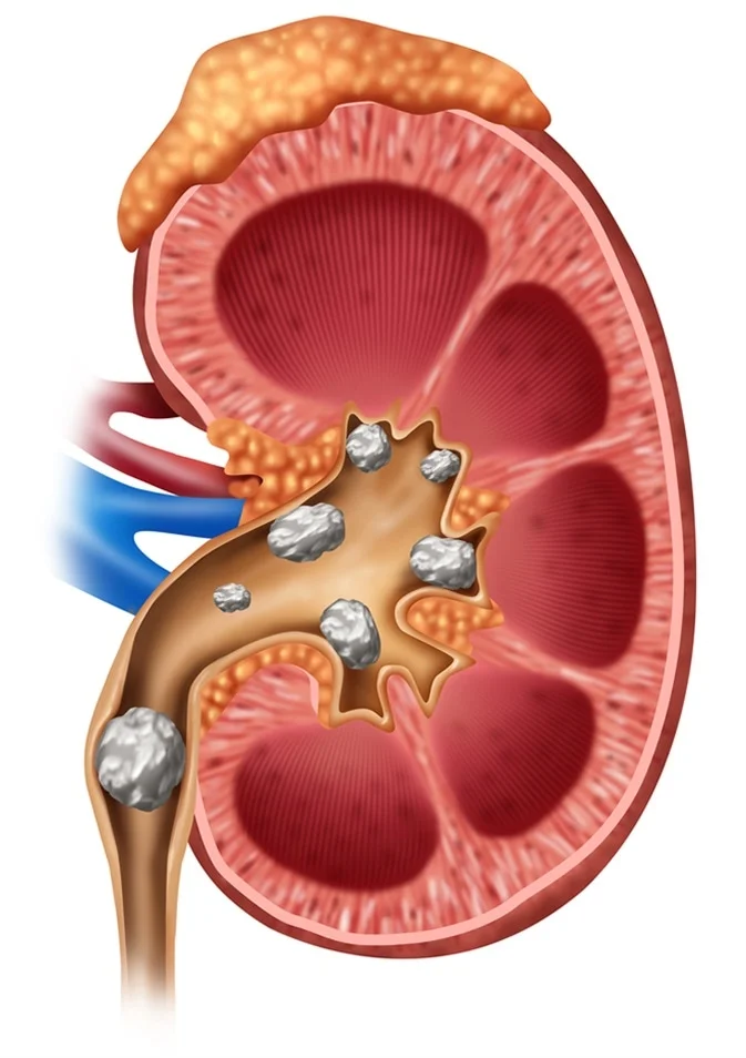
The results of the stone analysis can provide valuable information for determining the cause of the stone formation and guiding treatment and prevention strategies. For example, different types of kidney stones (such as calcium oxalate stones, uric acid stones, struvite stones, and cystine stones) have distinct compositions and may require different dietary and medical interventions for prevention.
If you’re interested in seeing images of kidney stones and their analysis, you can search online for medical resources or consult with a healthcare provider who may be able to provide relevant educational materials.

Reviews
There are no reviews yet.