Description
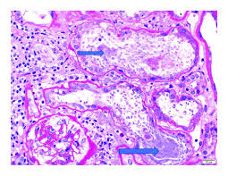
Histopathology of kidney biopsy Panel 2 refers to a specific set of tests and examinations performed on a kidney tissue sample obtained through biopsy. Histopathology involves the microscopic examination of tissues to study the changes or abnormalities at the cellular level.
Panel 2 typically includes a range of stains and tests aimed at identifying various structures and abnormalities within the kidney tissue sample. The specific components of Panel 2 can vary depending on the laboratory and the clinical indication for the biopsy. However, some common components may include:
- Hematoxylin and eosin (H&E) staining: This is a standard staining method used to visualize cellular structures and general tissue architecture. It helps identify abnormalities such as inflammation, fibrosis, and cellular changes.
- Periodic acid-Schiff (PAS) staining: PAS staining highlights glycogen, mucosubstances, and basement membranes in tissues. It can help identify features such as glomerular basement membrane thickening in conditions like diabetic nephropathy.
- Trichrome staining: Trichrome stains, such as Masson’s trichrome, are used to visualize collagen fibers and assess fibrosis within the kidney tissue. Fibrosis is a common feature of chronic kidney disease.
- Immunofluorescence (IF) or Immunohistochemistry (IHC): These techniques involve using specific antibodies to detect the presence and location of proteins or antigens within the tissue sample. Immunofluorescence is commonly used to identify immune complex deposits in conditions like lupus nephritis, while immunohistochemistry can help identify specific cell types or markers.
- Electron microscopy (EM): In some cases, electron microscopy may be performed to examine ultrastructural features of kidney tissue, such as podocyte foot process effacement in nephrotic syndrome or the presence of electron-dense deposits in glomerular diseases.
These tests and stains provide valuable information about the underlying cause and severity of kidney disease, guiding diagnosis, prognosis, and treatment decisions. The interpretation of histopathological findings requires expertise from pathologists specialized in renal pathology.

Reviews
There are no reviews yet.