Description
Histopathology of a small biopsy specimen involves examining tissue samples under a microscope to identify any abnormalities or diseases. Here’s an overview of the process:
Procedure:
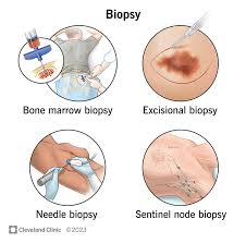
- Biopsy Collection: A small tissue sample is taken from the body, often using a needle or during a surgical procedure.
- Fixation: The tissue sample is preserved in a fixative solution (usually formalin) to prevent decay and maintain cellular structure.
- Processing: The fixed tissue is processed in the laboratory, where it is dehydrated, embedded in paraffin wax, and sliced into thin sections.
- Staining: Sections of the tissue are mounted on glass slides and stained with dyes (such as hematoxylin and eosin) to highlight different structures and cell types.
- Microscopic Examination: A pathologist examines the stained tissue sections under a microscope to assess the cellular morphology, architecture, and any abnormalities present.
Purpose:
- Diagnosis: Histopathology helps diagnose various conditions, such as cancer, inflammatory diseases, infections, and structural abnormalities.
- Prognosis: It provides information about the aggressiveness of tumors or the extent of disease progression.
Reporting:
- Pathology Report: A detailed report is generated with findings describing the tissue’s characteristics, any abnormalities detected, and the pathologist’s interpretation.
Follow-Up:
- Treatment Planning: Histopathology results guide treatment decisions, such as surgery, chemotherapy, radiation therapy, or further diagnostic tests.
- Monitoring: Periodic biopsies may be necessary to monitor disease progression or treatment effectiveness.
Histopathology is crucial for accurate diagnosis and plays a significant role in personalized medicine by tailoring treatments to individual patient needs based on tissue characteristics.

Reviews
There are no reviews yet.